
The MRI Was Normal. The Lesion Was Still There.
 FND Nope
Moderator
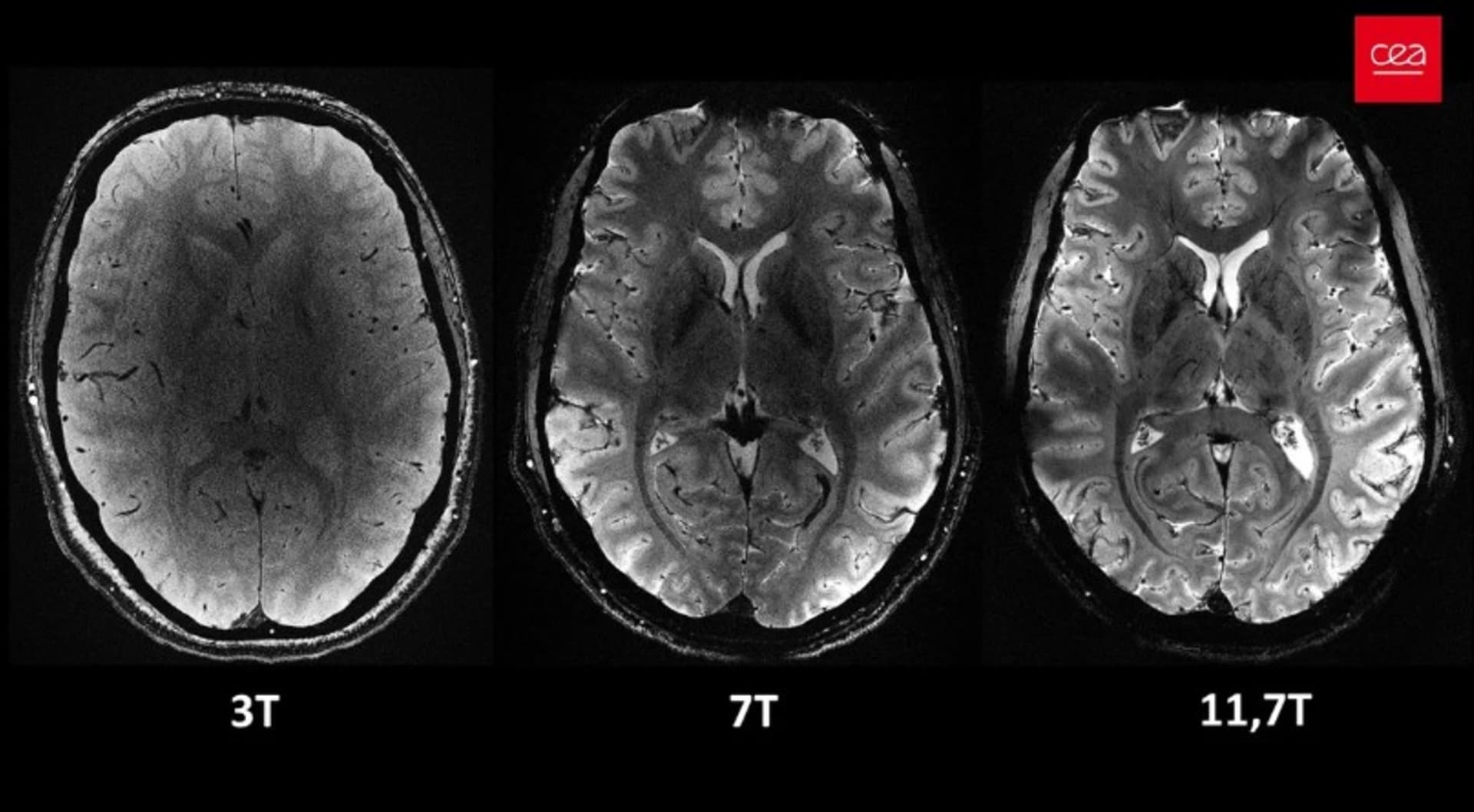
Apparently, a normal MRI does not always mean there is nothing wrong with the brain. Sometimes it means the scanner simply could not see what was there. That is not speculation. Researchers using ultra-high-field 7 Tesla MRI have identified structural abnormalities that conventional scans failed to detect, including lesions subsequently confirmed through surgery and examination of brain tissue.
Meanwhile, the argument that not every neurological patient needs an MRI continues to appear in discussions about Functional Neurological Disorder (FND). Technically, that statement is true. Not everyone needs imaging. But it becomes a rather different proposition when diagnostic confidence is used to dismiss further investigation in a patient whose symptoms remain unexplained, progress, or fail to fit the original diagnosis. The evidence now gives us an excellent reason to question that confidence.
When a Normal MRI Wasn't Normal After All
A study published in Epilepsia Open examined 40 patients with drug-resistant focal epilepsy whose previous lower-field MRI scans had been normal or had failed to explain their seizures. Researchers used 7T MRI and identified previously unrecognised lesions in nine patients, approximately 23% of the group. Six underwent surgery, and examination of the removed tissue confirmed focal cortical dysplasia or related abnormalities of cortical development.
Read that again. These patients had undergone conventional imaging, yet abnormalities were subsequently identified using more advanced technology and confirmed through histopathology. This was not simply a radiologist spotting a suspicious shadow and declaring victory. There was physical evidence of abnormal brain tissue.
The study involved a carefully selected group of patients with refractory focal epilepsy, not an unselected population with neurological symptoms. Its findings therefore cannot be used to claim that 23% of all patients with normal MRI scans have hidden lesions. What it establishes is narrower, but devastating to any claim that normal conventional imaging excludes all structural pathology: in some patients, it demonstrably does not.
Sixteen Patients, Conventional MRI Negative, Seven With Potentially Epileptogenic Findings
A separate study published in Epilepsy & Behavior Reports investigated 16 patients with focal epilepsy whose previous 1.5T or 3T MRI scans had been classified as non-lesional. When researchers examined them using 7T MRI, seven had findings judged to have definite epileptogenic potential in the context of their clinical and surgical evaluations.
All 16 patients underwent neurosurgical intervention. Among the seven with definite 7T findings, four achieved freedom from disabling seizures, compared with three of the nine patients without definite findings. The researchers emphasised that 7T findings were interpreted alongside other clinical and electrophysiological evidence, not used independently to justify surgery.
One particularly striking example involved polymicrogyria, an abnormality of cortical development, which was not detected on the earlier 1.5T images but was clearly visualised at 7T. The paper includes comparative images. The difference was not whether the patient had an abnormality on Monday but developed one on Tuesday. The difference was what the imaging technology could reveal.
Multiple Sclerosis: Even Ordinary MRI Upgrades Find More Lesions
This problem is not confined to epilepsy. A multicentre study published in Neurology compared 1.5T and 3T MRI in patients presenting with clinically isolated syndrome, a neurological episode potentially suggestive of multiple sclerosis. The stronger 3T scanners detected 15% more T2 brain lesions than 1.5T scanners, with increased detection in several anatomical regions.
Interestingly, the additional lesions did not result in more patients meeting the diagnostic criteria for MS during the study. That distinction matters. Detecting additional pathology and changing a formal diagnosis are not the same thing. Nevertheless, the central observation remains: the same patients, examined with different imaging capabilities, produced different lesion counts.
So when someone announces that a scan is normal, it is reasonable to ask which scanner was used, which anatomical regions were examined, what imaging sequences were performed, and what abnormalities those techniques were capable of detecting. A radiology report is an interpretation of the examination performed. It is not a certificate guaranteeing the absence of every possible neurological abnormality.
Source: Three-Tesla MRI does not improve the diagnosis of multiple sclerosis: A multicenter study.
And 7T MRI Finds Even More MS Lesions
A 2021 systematic review published in NeuroImage: Clinical found that 7T MRI detected, on average, 52% more cortical MS lesions than the best-performing 3T imaging sequences in the studies compared. The improvement was particularly notable for lesions within the cerebral cortex, which are notoriously difficult to visualise using conventional MRI.
Here is the part that should make anyone uncomfortable with excessive diagnostic certainty: even 7T MRI failed to detect all lesions when compared with post-mortem pathological examination. In other words, researchers could examine actual brain tissue and find abnormalities that even ultra-high-field imaging had not reliably identified.
The technology is improving, but its limitations have not magically disappeared. A negative scan is evidence about what an examination detected, not an absolute statement about everything that might exist within the nervous system.
Source: Imaging cortical multiple sclerosis lesions with ultra-high field MRI.
And Yet, Apparently, Not Everyone Needs an MRI
Now we arrive at the rather extraordinary contradiction in the way neurological investigations are sometimes discussed. Researchers are demonstrating that progressively more sophisticated MRI technology can reveal previously undetected structural pathology, while arguments about avoiding imaging in patients suspected of having FND continue to circulate.
To be precise, not every patient presenting with neurological symptoms requires an MRI. Imaging decisions depend on the symptoms, examination, suspected diagnoses and clinical circumstances. Nor is 7T MRI currently a routine investigation for every neurological complaint. But those qualifications do not excuse using diagnostic confidence as a reason to ignore evidence that further investigation may be necessary.
In fact, a 2024 review published in Brain, specifically examining iatrogenic harm in FND, warned about diagnostic overshadowing and under-investigation. The authors acknowledged that coexisting neurological and medical conditions can be overlooked, and that investigations are usually an essential part of assessing patients diagnosed with FND.
That is worth emphasising. Even literature written from within the FND clinical field recognises the danger of allowing the diagnosis to interfere with investigation of other diseases. This is not an argument invented by critics. It is an acknowledged patient-safety problem.
Source: Iatrogenic harm in functional neurological disorder, Brain.
Another review, published in Clinical Medicine in 2021, explicitly identifies reliance on normal investigations and failure to consider coexisting medical conditions as diagnostic pitfalls. It also states that many neurological conditions may have normal structural imaging.
So the question is not whether every patient should be pushed through an MRI scanner. The question is whether the decision not to investigate has been justified by the patient's actual clinical presentation, or whether the diagnostic label has effectively ended the conversation.
Source: A practical review of functional neurological disorder for the general physician.
The Questions That Should Be Asked
Before a clinician treats negative imaging, or the decision not to obtain imaging, as sufficient reassurance against structural neurological disease, there are some fairly obvious questions worth answering:
- What was actually excluded? A normal brain MRI cannot exclude every spinal, peripheral nerve, neuromuscular, metabolic or genetic disorder. Nor can it exclude every microscopic abnormality within the brain.
- What imaging was performed? Was it 1.5T or 3T? Which sequences were used? Were the relevant anatomical regions examined? Was the protocol appropriate for the suspected condition?
- What happens when symptoms progress? Does new weakness, deterioration, a changing neurological examination or a new clinical finding trigger reassessment, or is everything interpreted through the existing diagnosis?
- Could two conditions coexist? Finding evidence supporting one diagnosis does not automatically establish that every symptom has the same cause.
- What evidence supports not investigating? Is there a documented clinical rationale, or is the decision based on confidence that the presentation is functional?
These are not unreasonable demands for exotic technology. They are basic questions about the scope of an investigation, the limits of diagnostic evidence and the need to reconsider conclusions when new information emerges. The importance of investigating possible comorbid disease is specifically addressed in the FND literature, while modern MS imaging guidance also recognises that higher-field MRI has limitations and that appropriate protocols matter.
Source: The use of 7T MRI in multiple sclerosis: review and consensus statement.
The Scanner Has Limitations. So Should Diagnostic Confidence.
None of these studies establishes that every person diagnosed with FND has an undiscovered lesion, or that upgrading every MRI would overturn the diagnosis. That would be an unsupported leap, and the research does not require one. What the evidence does establish is that clinically important structural abnormalities can remain undetected on conventional imaging, and that newer imaging methods can sometimes reveal them.
That should be enough to make any clinician cautious about treating a normal scan as definitive proof that structural disease is absent. It should also raise serious questions whenever an existing diagnostic label becomes a reason not to reconsider a patient's symptoms, especially when the clinical picture changes.
A normal MRI means no relevant abnormality was identified by that examination. It does not mean medicine has inspected every neuron and issued the brain a certificate of innocence.
The uncomfortable reality is that medical imaging continues to improve precisely because existing methods cannot reveal everything. The 7T studies demonstrate that this limitation is not merely theoretical. Previously undetected abnormalities have been found, investigated and, in selected cases, confirmed through surgery.
So when someone confidently announces that a neurological patient does not need imaging, perhaps the appropriate response is not automatic agreement or automatic outrage. Perhaps it is a much simpler question: What clinical evidence justifies that decision, and what would make you reconsider it?
Because the ability to recognise when a diagnosis needs to be revisited is not a weakness in clinical reasoning. Refusing to revisit it despite contradictory evidence is.