Diagnosed With Functional Seizures After Bereavement. It Was Autoimmune Encephalitis.
 FND Nope
Moderator
A woman in her 40s developed involuntary jerking movements shortly after losing a close family member. She had been experiencing depression, and her symptoms appeared to worsen with emotional stress. Doctors initially treated her episodes as psychogenic non-epileptic seizures (PNES), with functional neurological disorder (FND) also appearing in her working diagnosis. Unfortunately, the explanation was wrong. Further investigation eventually identified anti-LGI1 autoimmune encephalitis, an immune-mediated neurological disease capable of causing seizures, cognitive changes, psychiatric symptoms and abnormal movements. The case was published in Cureus on 8 October 2025.
The significance of this case is not simply that another neurological disease was initially mistaken for a functional disorder. It is how readily a recent emotional event became part of the explanation for progressively worsening neurological symptoms, while the underlying disease remained unidentified. The patient was not experiencing harmless stress-related movements. She had an inflammatory neurological condition requiring immunotherapy and antiseizure treatment.
From bereavement to a functional diagnosis
The patient had a history of hypothyroidism and hypertension but had reportedly been doing well until she developed depressive symptoms following the death of a close family member. She was initially managed with antidepressant medication. Approximately one month later, she began experiencing involuntary jerking movements affecting her left arm and leg. These episodes initially occurred once or twice daily but rapidly increased to between 20 and 30 episodes per day over just three to four days. Emotional stress made the movements worse, although she remained conscious during the episodes.
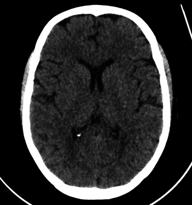
Her condition then escalated. While asleep, she experienced a generalized tonic-clonic seizure lasting approximately five minutes, witnessed by her son. The seizure involved oral secretions and was followed by confusion and memory loss lasting approximately 30 to 40 minutes. She was taken to an emergency department, where a CT scan of her head was reported as normal. A routine electroencephalogram (EEG) was also unremarkable. She was not started on antiseizure medication at that stage.
Despite the escalating movements and witnessed convulsive seizure, her working diagnosis included a functional disorder, involuntary jerks or a single isolated nocturnal convulsive seizure. The authors specifically describe how the patient's emotional stressor and earlier clinical encounters contributed to her being treated for PNES. A normal routine EEG does not rule out epilepsy, and a normal CT scan does not exclude autoimmune encephalitis. Neither result established that her movements were functional.
There is an important distinction here: PNES refers to seizure-like episodes that are not caused by epileptic electrical activity and is commonly classified within FND. But the absence of an abnormality on a routine EEG is not, by itself, proof of PNES. In this case, the patient's episodes were eventually linked by the authors to an autoimmune neurological disease.
The neurological evidence
An MRI scan revealed white matter hyperintensities and cortical thickening involving the right medial temporal lobe. These findings were not consistent with simply dismissing the presentation as psychological distress. When the patient sought a second neurological opinion because her movements persisted alongside anxiety and personality changes, she continued experiencing involuntary jerks during the examination. She was admitted for further investigation and started on levetiracetam, an antiseizure medication.
Her neurological examination was otherwise relatively unremarkable. Muscle strength, reflexes, sensation, gait and cranial nerve examination were normal. This is another important feature of the case: substantial neurological disease was present despite many conventional examination findings being normal.
Further investigations revealed several findings supporting autoimmune encephalitis:
- Anti-LGI1 antibodies: Positive at a titre of 1:1,000, providing strong evidence of LGI1-associated autoimmunity in the clinical context.
- Brain MRI: Abnormal signal and swelling in the right medial temporal lobe, a region commonly affected in limbic encephalitis.
- Involuntary movements: Frequent jerking episodes consistent with the movement abnormalities associated with the disease.
- Seizures: A witnessed generalized tonic-clonic seizure accompanied by prolonged post-seizure confusion and amnesia.
- Cognitive and behavioural changes: Memory difficulties, anxiety, mood changes and alterations in personality.
- Hyponatremia: Low blood sodium, identified by the authors as another feature supporting the diagnosis, although the case report does not provide a corresponding sodium value in its laboratory table.
The diagnosis was anti-LGI1 autoimmune encephalitis, a form of autoimmune limbic encephalitis involving antibodies against leucine-rich glioma-inactivated protein 1. This condition is associated with seizures, memory impairment, behavioural changes and characteristic involuntary movements. Some patients develop faciobrachial dystonic seizures, which can be brief, frequent and easily misinterpreted, particularly when routine EEG recordings do not capture clear epileptic activity.
The authors explicitly acknowledge the earlier diagnostic mistake:
"Till then, she was being treated for a functional neurological disorder"
That sentence deserves attention. The diagnosis changed because further neurological investigation produced evidence of a disease that had not been identified during the initial assessment. Her emotional history had not explained the movements. Her normal initial tests had not excluded neurological disease. The subsequent MRI findings and antibody results fundamentally changed the clinical picture.
Treatment revealed the seriousness of the disease
Once autoimmune encephalitis was confirmed, treatment changed substantially. The patient received intravenous methylprednisolone at 1 gram daily for four days, together with intravenous immunoglobulin (IVIG) over approximately four to five days. Levetiracetam was continued to control seizures. Her involuntary movements decreased, her mood improved and no further seizures were observed during the initial treatment period. She was discharged with a reported resolution of symptoms and plans for further immunotherapy.
However, the improvement was not the end of the story. At a four-week follow-up, she still had mild symptoms and remained positive for anti-LGI1 antibodies. She subsequently received two doses of rituximab, administered two weeks apart, with further improvement in symptom control. Approximately one month later, she returned to the emergency department after four generalized tonic-clonic seizures accompanied by intermittent absence-type episodes, as described by the authors.
A repeat MRI showed no significant change, but her EEG now demonstrated intermittent sharp-wave discharges in the right temporal region. Lacosamide was added to levetiracetam, and her seizures responded well. Her anti-LGI1 antibody titre had fallen to 1:10, and clinicians did not administer additional biological immunosuppression at that point. She was discharged on two antiseizure medications and remained seizure-free at the next reported follow-up one month later.
This was not a patient whose symptoms simply disappeared after reassurance or psychological intervention. She required high-dose corticosteroids, IVIG, rituximab and two antiseizure medications to manage an objectively identified neurological disease. Even after substantial initial improvement, she experienced further seizures requiring emergency treatment.
What this case exposes
The authors describe how autoimmune encephalitis can initially resemble psychiatric or functional conditions, particularly when behavioural symptoms, depression, anxiety or emotional stress accompany the neurological presentation. Anti-LGI1 encephalitis is especially important because its early manifestations can include brief abnormal movements, memory problems and personality changes before the full clinical picture becomes obvious. The disease is potentially treatable, but delayed recognition can mean delayed immunotherapy and continuing neurological deterioration.
The case also illustrates the danger of treating psychological context as diagnostic evidence. Bereavement is not a neurological investigation. Emotional stress exacerbating a movement does not establish its cause. Normal routine investigations do not automatically transform unexplained neurological symptoms into a functional disorder. When symptoms are progressing, the explanation needs to account for that progression rather than relying on the presence of an emotional event.
There are limits to what this report establishes. It describes one patient, not a measured rate of FND misdiagnosis. The authors retrospectively attribute her earlier PNES-like episodes to autoimmune encephalitis, but the report does not provide video-EEG confirmation of the precise mechanism of every early event. It nevertheless documents a patient initially treated for PNES/FND whose subsequent investigations established anti-LGI1 autoimmune encephalitis.
The most troubling aspect is how much had to happen before the correct disease was identified: rapidly escalating involuntary movements, a witnessed convulsive seizure, cognitive and behavioural changes, abnormal MRI findings and finally positive autoimmune antibody testing. The patient needed a second neurological opinion and additional investigation before treatment was directed at the underlying disease.
The bottom line: This woman was initially treated for a functional disorder while she had autoimmune encephalitis. Her recent bereavement helped shape the early interpretation of her symptoms, but it did not explain the neurological disease. The correct diagnosis emerged through further investigation, not through a better psychological explanation. That is precisely why progressive neurological symptoms should not be written off simply because a patient has experienced emotional distress.
References
- Longi AA, Fazlani M, Akram S, Nouri K, Shetty P. (2025). Autoimmune Encephalitis With Positive Anti-Leucine-Rich Glioma-Inactivated 1 (Anti-LGI1) Antibody Mimicking Psychogenic Non-epileptic Seizures. Cureus, 17(10), e94129. DOI: 10.7759/cureus.94129.
- Dash D, Tripathi M, Ihtisham K, Tripathi M. (2016). LGI1 encephalitis: a disease of jerks and confusion. BMJ Case Reports.
- Karvigh SA, Salehizadeh S, Vahabizad F. (2022). Anti-leucine-rich glioma-inactivated 1 encephalitis: two case reports and a review of the literature. Journal of Medical Case Reports, 16, 409.
- Endres D, Prüss H, Dressing A, et al. (2020). Psychiatric Manifestation of Anti-LGI1 Encephalitis. Brain Sciences, 10(6), 375.
- Ishikawa R, Sugimoto T, Ohno N, Iizuka T, Nomura E. (2025). A Case of Anti-N-Methyl-D-Aspartate Receptor (NMDAR) Encephalitis With Video-Documented Psychogenic Nonepileptic Seizures (PNES)-Mimicking Episodes Initially Considered as Somatic Symptom Disorder. Cureus.